
CT X-ray Equipment
SOLIO X Series
Superb quality and cost performance of SOLIO X Series has been paired with the proprietary software NEOPREMIUM2 as an integrated solution, offering uncompromising performance and comfort to the dental diagnosis environment.
Features
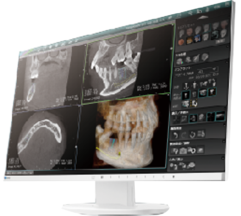
■ Variety of CT image reconstruction functions are available that are simple to access after CT image acquisition, which offers further diagnostic opportunities.
■ Incorporates many more helpful functions such as Tomosynthesis or dental image extraction from Panoramic images.
*Available when our software “NEOPREMIUM2” is used
- Quick and high quality image acquisition
-
Just 12 seconds of image acquisition for both 360-degree full scan CT exposure and standard Panoramic exposure.
Adding to the high-precision exposure, an array of image processing options are embedded such as CT image reconstruction or Tomosynthesis, to ensure both high quality images and quick acquisition.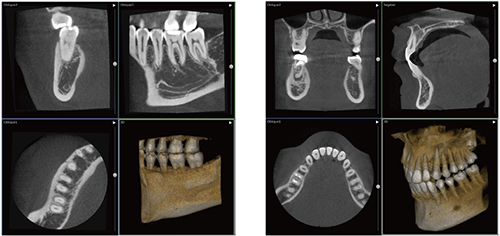
- CT
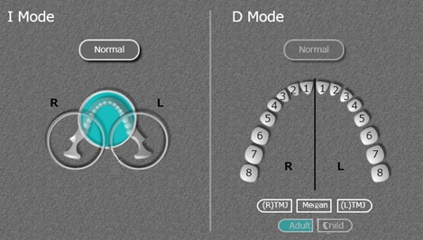
- Two exposure modes, D-Mode and I-Mode are incorporated.
12 seconds for high-precision CT images
360-degree rotation full-scan CT images take 12 seconds, and 180-degree CT images take 6 seconds. These are the quickest image acquisition time in our company history, all while precisely maintaining image quality.

360-degree Rotating Exposure for clear image quality
To reduce artifacts caused by metal objects, an exposure method using 360-degree rotation has been introduced. A larger exposure angle sweep works for minimizing the impact of imaging artifacts, so that sharp, high-precision CT images can be obtained.
**A rotation angle is automatically set to 270 degrees depending on the positioning of the patient, when the 360-degree rotation option is selected.CT image reconstruction function
After CT image is taken, various image reconstruction tools are available, all with simple operation.
■ NEODYNA MAR (Metal Artifact Reduction) *Optional
Our unique MAR algorism can reduce image artifacts due to metallic implant or other treatments. Combining with 360-degree imaging style can further realize with reduced impact from artifacts.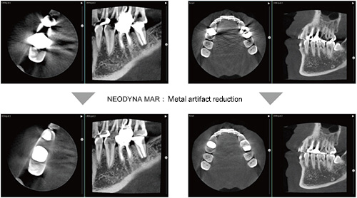
■ NEOSMART *Standard function
・ Sharp
Sharpens the image.・ Smooth
Smooths out the image.・ Scattered Ray Correction
Stabilizes the brightness of the hard tissues.・ Beam Hardening Correction
Reduces artifacts between implant fixtures.Part Reconstruction Function
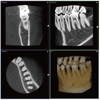
The obtained CT images can be reconstructed into an image with a minimum voxel size of 0.075 mm using D-mode and I-mode, within the range of ɸ40mm×50mm(H).
Without re-exposure, a high-precision image of the region of interest can be obtained.
*In the I-Mode, a voxel size of 0.1 mm, within the range of ɸ51mm×54mm(H) can be set.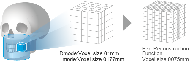
- 2D
-
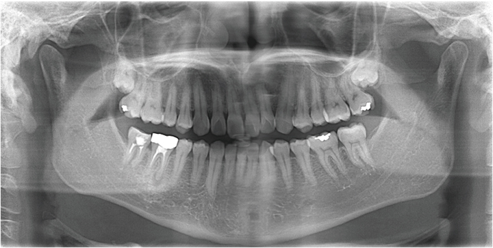
Especially committed to the panoramic image quality, we have added our unique image processing techniques for super-high precision images.

Tomosynthesis
Acquisition of Panoramic images in Tomosynthesis mode provides image data with a slice depth of 30mm, enabling a clear view of the blurring of the anterior teeth image area by choosing an optimum slice position.
*For images of juveniles or orthographic images, the acquisition area of panoramic image data is different.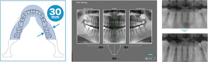
■ Automatic display can be performed with optimal slice positioning for the anterior teeth, from a region with a slice depth of 30 mm.
■ It is possible to adjust the orbit of panoramic images for each of the anterior teeth and the left and right molars to obtain a set of images best matching the shape of the patient’s dentition.
*Changing and saving the panoramic image will overwrite the image with the changed orbit. Before saving, ensure that the panoramic image displayed on the screen is the image you wish to save.■ Clearer images can be displayed using data from 31 images spaced at 1 mm intervals.
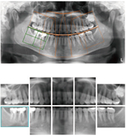
Dental Image Extraction
●Dental images can be extracted using the 10-image/14-image methods.
●Configuration of the region to extract or image modification is available.
Increased Quality in Panoramic Images
Frequency processing and noise reduction supported by our original image processing technology have realized higher image quality.
This can suppress graininess often seen at temporomandibular joint and molar region, helping to diagnosis of caries and inflammation.
- Comfortable exposure environment
-
Rich and supportive functions including a new head support & grip, CT position systems have realized a comfortable exposure environment that minimizes burdens both on patients and operators.
Easy and comfortable CT exposure area setting
Easy operation allows CT exposure without burdens on patients and operators.
-
Automatic Exposure Area Setting
The PC operation can select the CT exposure area for automatic movement of the equipment to the set position, allowing horizontal positioning before a patient is introduced.
-
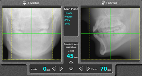
Preliminary Imaging Function to accurately set the CT exposure area
The imaging position can be corrected for all directions; front/back, left/right, up/down of the CT exposure area, enabling to set the CT exposure area accurately and reliably. After setting, the exposure mechanism automatically moves to the correction position, for a correct exposure of the target diagnostic area. This prevents a need of re-exposure due to a mistaken area setting.
CT Positioning System
After a patient is positioned, the exposure area can be vertically aligned without asking to move patients, which can drastically minimize the burdens on both patients and operators.
Unique head support & grip to enable Panoramic image acquisition in correct positioning
Adding to the head part, securely fixing at two chest parts (totally 7-point support) allows Panoramic image acquisition without blurring in a correct patient position with the cervical spine stretched.
Also, minimizing the needs of parts replacement according to the exposure mode also enables smooth positioning.
*Some body shapes of patients are not supported by the fixture at the chest part.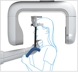
-
- Space-saving, Secure & Safe Operation
-
Installation in a restricted space, a solution of permanent secure and safe operation.
-
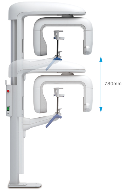
780mm stroke supporting both standing and sitting positions
Vertical stroke range of 780mm covers wide operation range including standing and sitting position and wheelchair use.
-
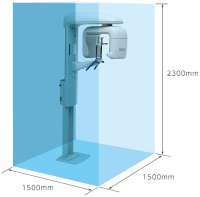
Space-saving, Compact CT Equipment
As the most compact CT equipment in our history, it can easily fit into an X-ray booth of 1500 mm width and depth.
Compact Sensor & Wide Arm
Reducing the size at the lower part of the sensor, adding to the wide-arm design, minimizes the possibility of the patient’s shoulder inadvertently touching the rotating part of the equipment.
-
Exposure mode
CT
 D-Mode: ɸ51mm×55mm(H) voxel size: 0.1mm
Enables high-resolution imaging of a localized area, for detailed observation of teeth and bones.
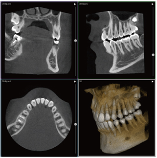 I-Mode: ɸ90mm×91mm(H) Voxel size: 0.177mm
Full arch images are available: one-shot allows the entire dentition area including the No.8 impacted apex.
2D
 Panoramic: 12 sec. (Normal)
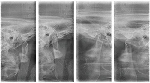 Lateral TMJ :3sec.(x4times)
CT X-ray Equipment
SOLIO X Series Catalog(PDF)
Catalog download
- Dental X-ray Products
