Philosophy

Imaging new visions
Medical care changes through the ability to observe.
It changes treatments, so that the sick can be returned to health.
More than anything, it can change a patient’s anxiety into hope.
From clearer images we enable higher quality treatment,
better treatment decisions, so advancing medical care.
Imaging new visions, for better medical care.
It is our mission and our responsibility to follow this path set out before us.
Image Quality
Going back to the time of analog devices, we have pursued and realized technology development in rotation orbits and X-ray output methods.
Now in the age of digital devices, we have further improved our image correction technology; improvements including the adjustment in the addition of pixel-shifting during panoramic image output, as well as further advancements in our original technology for optimizing parameters in the tone curve to stabilize the shading of the images.
Since the advent of CT scanning, image artifacts arising from metal objects interfering with the X-rays has become one of the major obstacles to diagnostic imaging. Our focus on the image processing technology has never stopped, which is well demonstrated in the development of the “Metal Artifact Reduction“ function which processes images more effectively by applying appropriate corrections to artifacts that vary by material.
The pursuit of ever clearer and more accurate images that contribute to better dental treatment. That is at the core of our technology development.
Usability
We focus not only on the image quality, but also on the processing speed and ease of use of our own software after capturing the image.
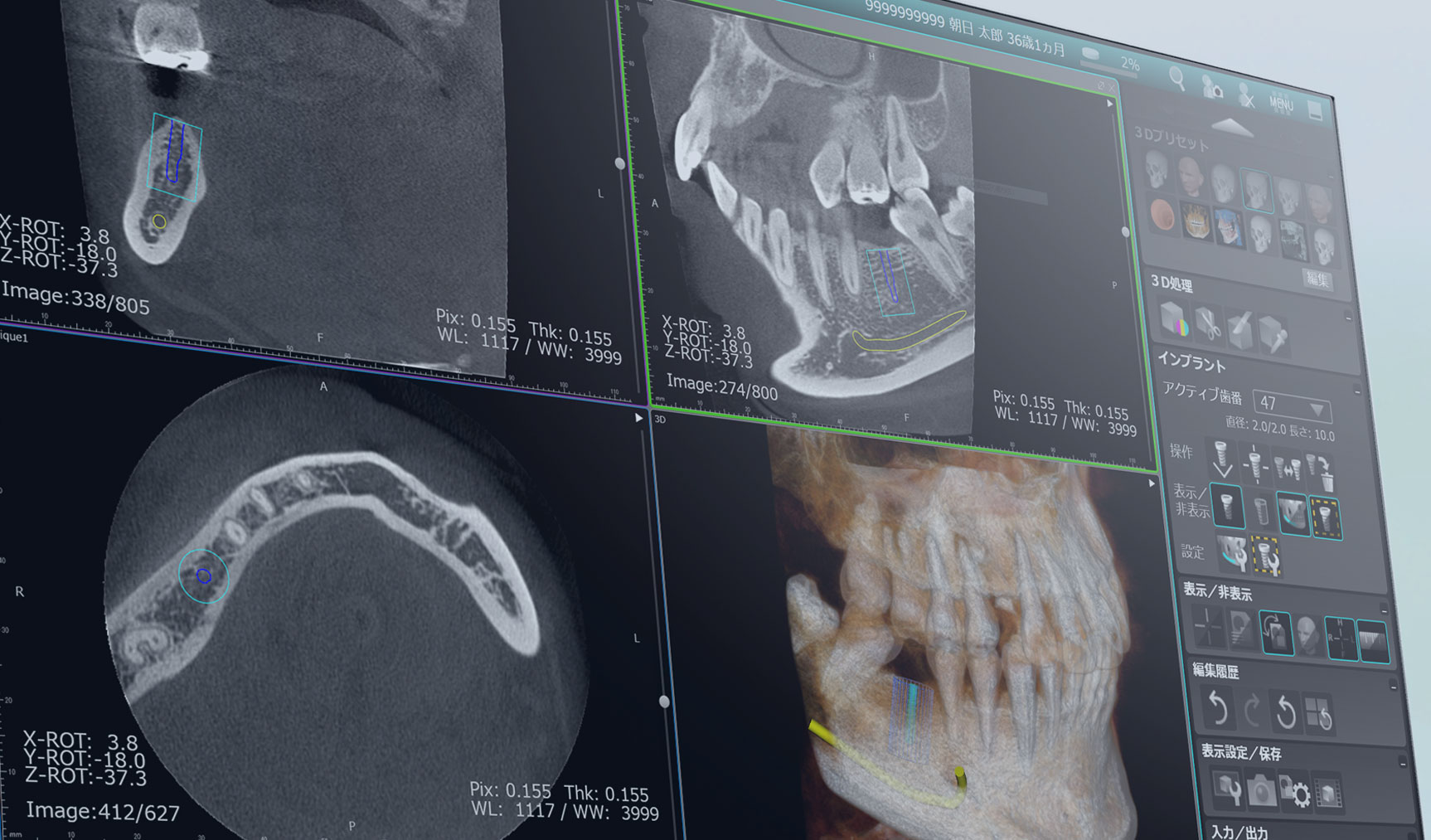
Using a GPU (Graphics Processing Unit) with specialized image processing for image creation processes and 3D graphics display, we have realized the image data handling in high speed needed to reduce wait time when every second counts, as well as the smooth rendering of 3D images.
On the screen for the patient to view, function buttons are summarized with icons at the right side of the screen in order to reduce the need to move the mouse, with the acquired image taking a large display area at the center of the screen. The screen is customized so that on the right side, only icons relevant to the image type appear (panoramic/dental/CT). This is a user interface designed to reduce operational errors and provide seamless explanations during dental treatment.

Comfort
No patients want to feel unnecessary stress when they come to dental clinics with pain and anxiety. That is why we make sure that our X-ray equipment has the specifications to give the smoothest experience while imaging.
The vertical motion range of the unit is ergonomically designed to be optimally positioned for both standing and seated positions, and for both children and taller people.
The vertical motion can be adjusted within approx. 780 mm on average, and once the patient is securely positioned, the imaging area can be smoothly and quickly adjusted by using the pre-scan function in 1 mm increments in all directions (front/back, up/down, left/right) either from the PC or the operation panel, without changing the patient’s posture at all.
By reducing a patient’s “physical pain” and “mental anxiety” caused by changing posture while the head is immobilized or any undue time spent on positioning, the system makes radiographic treatment more comfortable also for the dentists.

Safety
X-ray safety is a concern for patients.
We have been continually pursuing functions to minimize the patient’s exposure dose.
In the CT equipment, after securely positioning a patient, the image capturing area in each direction (front/back, up/down, right/left) can be finely adjusted in 1 mm increments without changing the patient’s posture. This feature allows the user to center the region of interest on any part of the image, which accordingly allows imaging of a smaller area, resulting in a 64% reduction in dose compared to imaging a larger area (internal comparison between I-Mode and D-Mode).
To realize clearer image quality, the 360-degree rotational imaging method is available to minimize artifacts due to metallic objects. Larger rotation angles reduce the effects of artifacts in images, and sharper, higher precision CT images can be obtained.
On the other hand, the 180-degree rotation option is also available, which halves the exposure time, so reducing the exposure dose by 50% compared to the 360-degree rotation option.
The availability of these options increases the choices for treatment and care, contributing to the optimum treatment regime for each patient case.
*In the 180-degree rotation scans, the amount of image noise is higher compared to larger rotation angles of 270° and 360°.
**A rotation angle is automatically set to 270 degrees depending on the positioning of the patient, when the 360-degree rotation option is selected.
