
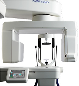
CT X-ray Equipment
AUGE SOLIO Series
Clear, high-precision image quality giving powerful diagnostic assistance, with excellent operability via the 5.7-inch touch panel.
AUGE SOLIO Series are all-in-one systems using our unique technologies and rich expertise. These models fulfill the large variety of needs required from diagnostic imaging for dental treatment.
For the increasing demands of CT imaging, particularly, we have realized precise diagnostic imaging with a wide exposure range and a patient-friendly, accurate positioning system in addition to its high-resolution exposure capability, which is one of the features of its dental cone beam CT.
Made in Kyoto and handed down over half a century, our craftsmanship delivers a renewed experience and hospitality.
Features
Clear, high-precision image quality giving powerful diagnostic assistance, with excellent operability via the 5.7-inch touch panel.
AUGE SOLIO Series are all-in-one systems using our unique technologies and rich expertise. These models fulfill the large variety of needs required from diagnostic imaging for dental treatment.
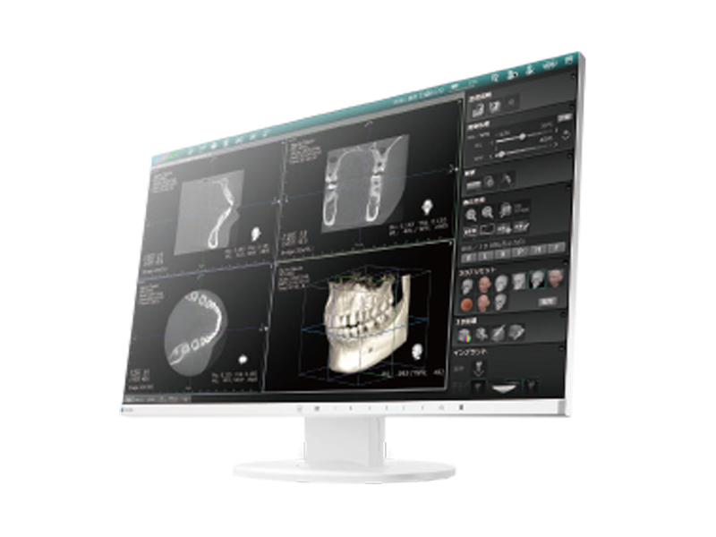
- CT image reconstruction function
-
After a CT image is taken, various image reconstruction functions are available with simple operation.
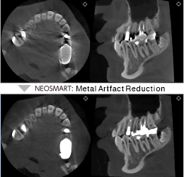
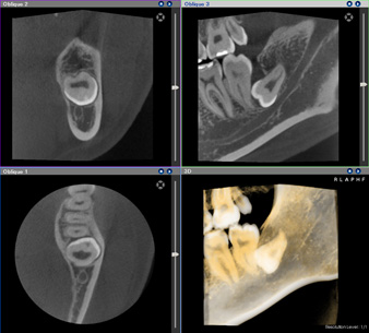
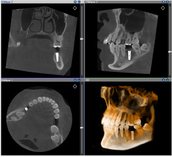
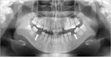
■ NEODYNA MAR (Metal Artifact Reduction) *Optional
Our unique MAR algorism can reduce image artifacts due to metallic implant or other treatments. Combining with 360-degree imaging style can further realize with reduced impact from artifacts.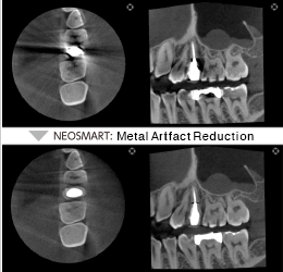
・ NEOSMART *Standard function
Sharpens the image.・ Smooth
Smooths out the image.・ Scattered ray correction
Stabilizes the luminance of dental hard tissue.・ Beam hardening correction
Reduces artifacts between implant fixtures
*Available functions depend on the exposure mode.
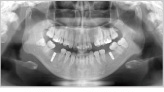
- Tomosynthesis Function
-
Acquisition of Panoramic images in Tomosynthesis mode provides image data with a slice depth of 30mm, enabling a clear view of the blurring of the anterior teeth image area by choosing an optimum slice position.
*For images of juveniles or orthographic images, the acquisition area of panoramic image data is different.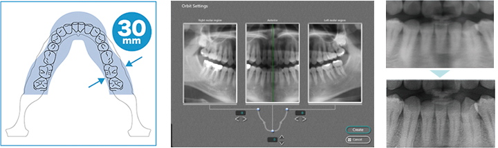
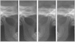
■ The optimum slice positions of the anterior teeth are automatically displayed from the image data with a slice depth of 30 mm.
■ The orbit of panoramic images can be adjusted for each of the anterior teeth and the left, right molars to obtain a set of images best matching the shape of the patient’s dentition.
*Changing and saving the panoramic image will overwrite the image with the changed orbit. Before saving, ensure that the panoramic image displayed on the screen is the image you wish to save.■ Clearer images can be displayed using data from 31 images spaced at 1 mm intervals.
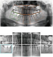
Dental image extraction
● Dental images can be extracted using the 10-image/14-image methods.
● Configuration of the region to extract or image modification is available.
- Wide Arm Design to eliminate image distortion
-
A wide-arm design means the transmitted X-rays are almost parallel to each other enabling high-precision images without distortion.
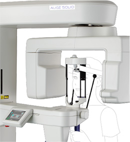
- Head Support System to prevent motion artifacts
- The head support securely holds a patient’s head without burdening it. It minimizes the impact to the images due to head movement, realizing a high-precision image.
- 360-degree Rotating Exposure to realize clear image quality
-
To reduce artifacts caused by metal objects, an exposure style of 360-degree rotation has been introduced. A larger exposure degree works for more minimized impact to the images due to artifacts, and a sharp, high-precision CT images can be obtained.
**A rotation angle is automatically set to 270 degrees depending on the positioning of the patient, when the 360-degree rotation option is selected.
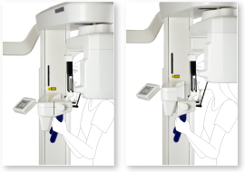
- CT Positioning System to enable vertical movement of the CT exposure area
-
After the patient is positioned, the exposure area can be vertically aligned without asking the patient to move, which can drastically minimize the burden on both the patient and the operator.
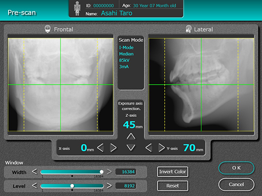
- Pre-scan Imaging Function to accurately set the CT exposure area
-
The imaging position can be corrected for all directions; front/back, left/right, up/down of the CT exposure area, enabling to set the CT exposure area accurately and reliably. After setting, the exposure mechanism automatically moves to an optimum position for a correct exposure of the target diagnostic area. This prevents the need of re-exposure due to a mistaken area setting.
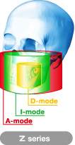
Exposure mode
There are multiple modes covering various exposure areas to support various cases
D-Mode to cover dentition / temporomandibular joint. I-Mode for the entire dentition area. Mode A to cover temporomandibular joint and both jaws. The optimum exposure mode can be selected according to the target dental treatment.
Exposure Mode (3D imaging)
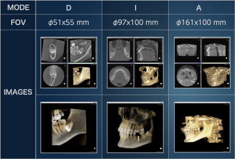
 D-Mode: ɸ51×55mm Voxel size: 0.1mm
 I-Mode: ɸ97x100mm Voxel size: 0.19mm
(Display of reproduction available at ɸ80x90mm, voxel size of approx. 0.15mm)
Exposure Mode (2D imaging)
 Panoramic (Normal): 12 sec.
 Panoramic (High-speed): 9 sec.
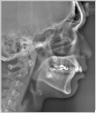
 Lateral TMJ: 3 sec. (x4)
 PA TMJ: 3 sec (x 2)
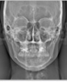
 Maxillary sinus: 8 sec.
Exposure Mode(CM type)
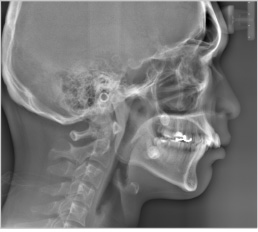 Cephalometric (Lateral, Normal): 4 sec.
 Cephalometric (Lateral, Short time): 2.9 sec.
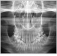
 Cephalometric (PA): 4 sec.
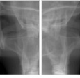
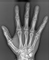 Bones of Carpus: 4 sec.

CT X-ray Equipment
AUGE SOLIO Series Catalog(PDF)
Catalog download
- Dental X-ray Products
