Product

Arm-type X-ray CT diagnostic instrument
SOLIO X Series
IMPROVED IMAGE QUALITY
By integrating a new sensor, even clearer panoramic and CT images are
now possible.
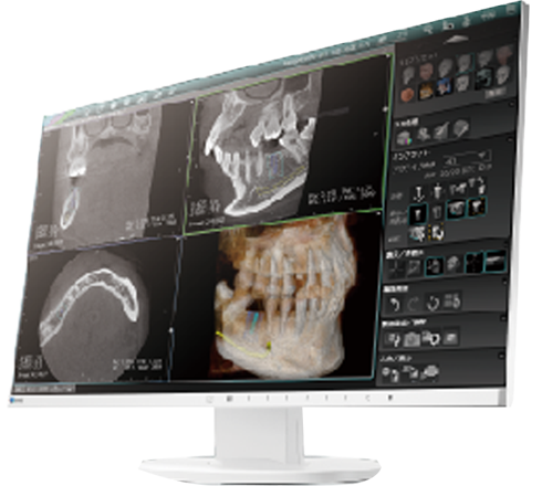
The obtained panoramic images are ideal for full-mouse screenings,
whereas the CT images, through 3D scanning, are for detailed diagnoses
of teeth and bones from all aspects.
Enhanced panoramic imaging, the foundation of dental treatment, combined
with high resolution CT imaging for detailed diagnosis, has transformed
the way we work. We can see more, diagnose with confidence, and truly
embrace Imaging New Visions.
*This video is in Japanese only.


PANORAMA

Integrated optimized sensor for panoramic images

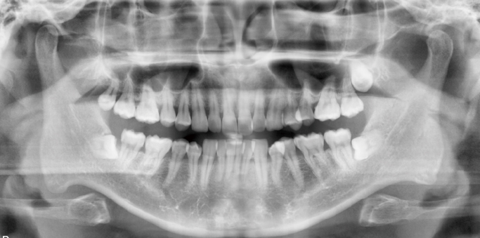
AF (Asahi Fine) Panorama
By combining the three features: Noise Reduction, Sharpening and
Focal Composition, our newly developed image processing algorithm
gives clearer images.
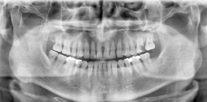
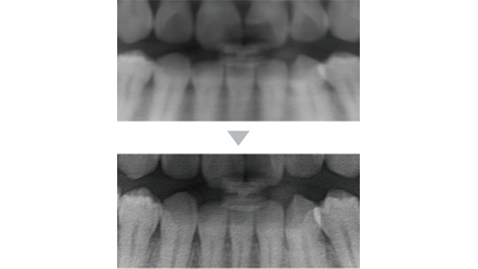
Focal Composition
Dual-image processing technology has been introduced for composite
panoramic images constructed from multiple tomographic planes to
enhance the clarity of the contours in any part of the image.
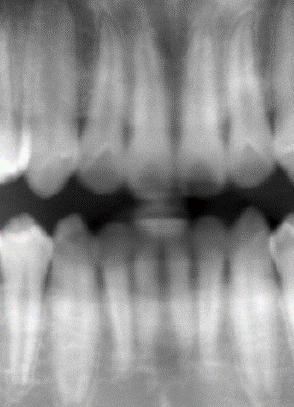
Normal panoramic images
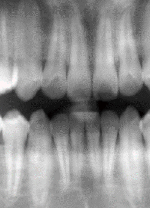
Focal Composition processed
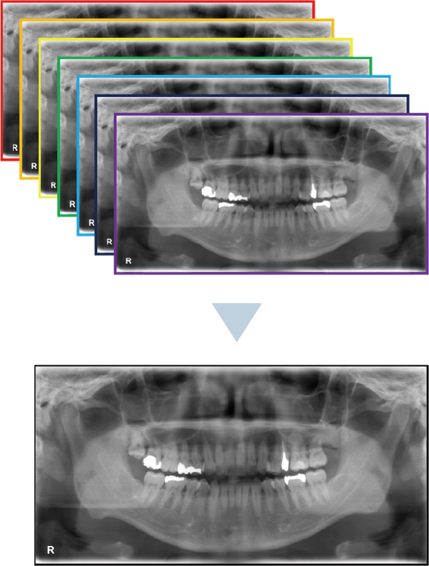
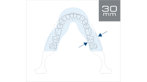
Tomosynthesis
The tomosynthesis mode allows panoramic images of a slice thickness
of 30 mm and optimizes the image by adjusting the blurred area at
the anterior teeth. As clearer images are always available, no
repeated exposure is required even after misaligned processes.
*Children need to have different focal areas for panoramic images.
Optimized slice position images are automatically displayed
from the image data of an area with a slice thickness of 30
mm.
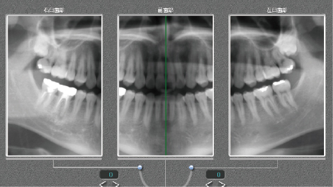
As the panoramic imaging trajectory can be adjusted in the
anterior and right and left molar regions, images can be
optimized according to the shape of the patient's dentition.
*Once saved, the trajectory cannot be corrected.
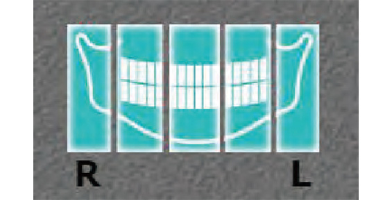
Clearer images can be generated from 31 images with a thinner
slice thickness of 1 mm.
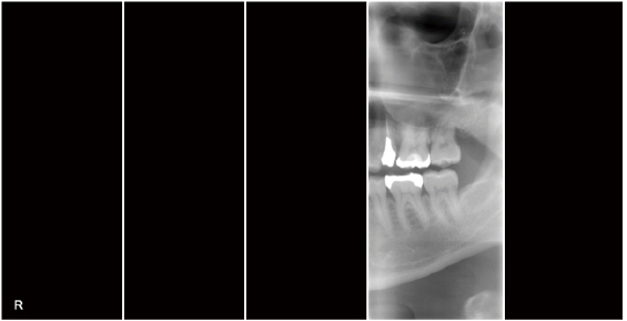
Partial panorama
Limiting exposure to specific areas is possible by eliminating
unnecessary radiation. This approach is particularly effective for
patients who experience difficulties when undergoing dental imaging
procedures due to involuntary choking reflexes or other reactions.
CT

High Definition CT Images Takes Only 12 sec
12 sec for 360-degree full-scan CT images, and 6 sec for 180-degree
CT images. Our shortest imaging time ever, without compromising
image quality.
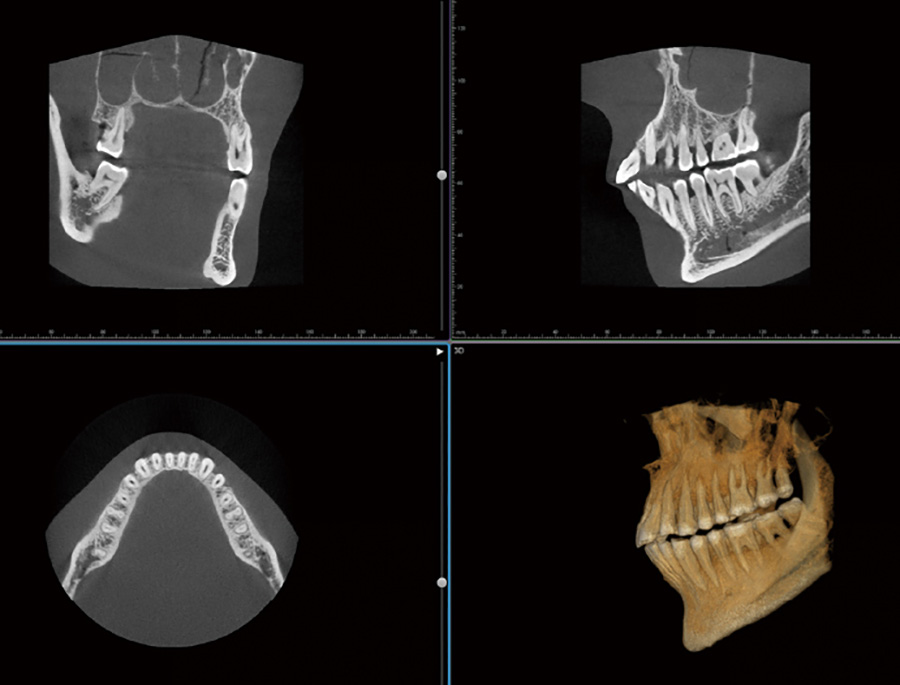
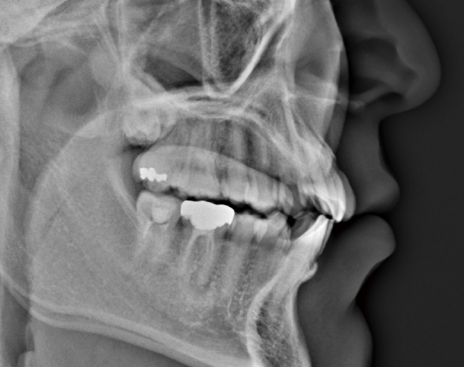
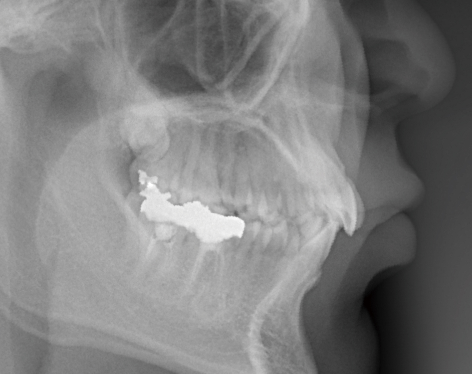
360-degree Exposure Realizing Clear Image Quality
To minimize image artifacts caused by the presence of metal, we have
introduced a 360-degree rotational shooting system. The larger the
rotation angle, the less impact on the image due to artifacts,
resulting in a crisp high-definition CT image.
*A rotation angle of 270 degrees is possible according to the
patient’s positioning.
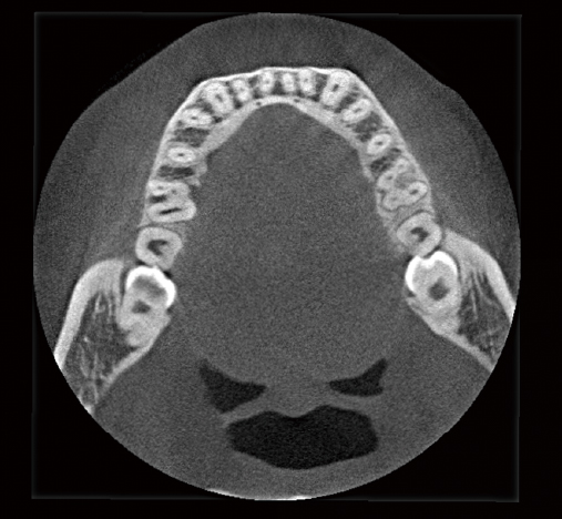
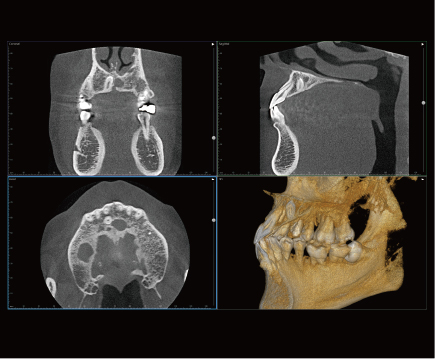
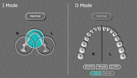
FOV (I-mode) coverage up to the third molar apex
Exposure Mode, Field of Vision (FOV)

D-mode
Ф51 × 55 (H) mm
Voxel size: 100 μm
Voxel size: 100 μm
I-mode
Ф98 × 100 (H) mm
Voxel size: 177 μm
Voxel size: 177 μm
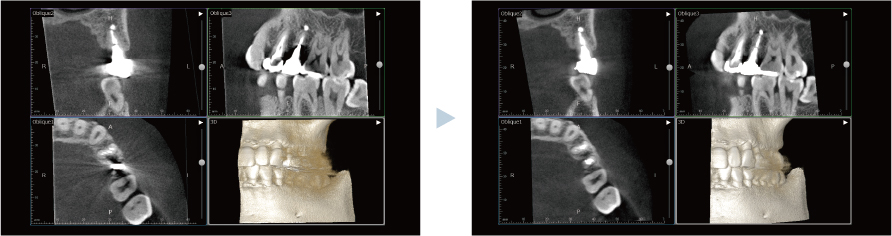
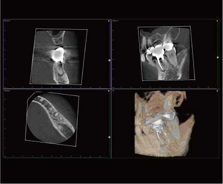
NEODYNA MAR (with reduced metal-induced artifacts)
Our proprietary MAR algorithm reduces image artifacts caused by
metal implants and other objects. As well as 360-degree image
acquisition, images can be obtained that are less affected by
artifacts.
NEOSMART
Sharpened - Scattered ray correction
Stabilizes the brightness values of hard tissues
Smooth - Beam hardening correction
Decreases image artifacts between tooth implants.
Part Reconstruction Function
Without the necessity of re-exposure, a high-precision image of the
region of interest can be obtained.
| Normal exposure | Part Reconstruction applied | |
|---|---|---|
| D-mode |
Ø51×55 (H) mm Voxel size: 100 μm |
Ø40×50 (H) mm Voxel size: 75 μm |
| I-mode |
Ø98×100 (H) mm Voxel size: 177 μm |
Ø51×54 (H) mm Voxel size: 100 μm |
|
Ø40×50 (H) mm Voxel size: 75 μm |
NEODYNA MAR / NEOSMART
*This function is available only in “NEOPREMIUM2”software
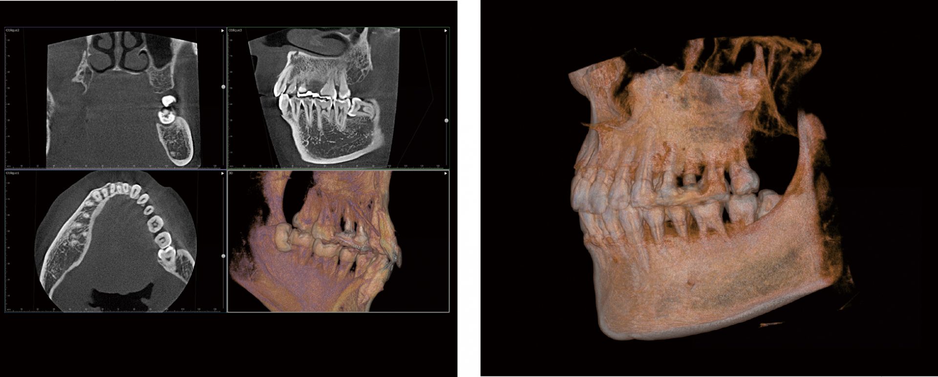
Gingival root (permanent maxillary right second molar),
impacted wisdom tooth
Apical periodontitis
Impacted supernumerary tooth

Enlargement of the periodontal cavity
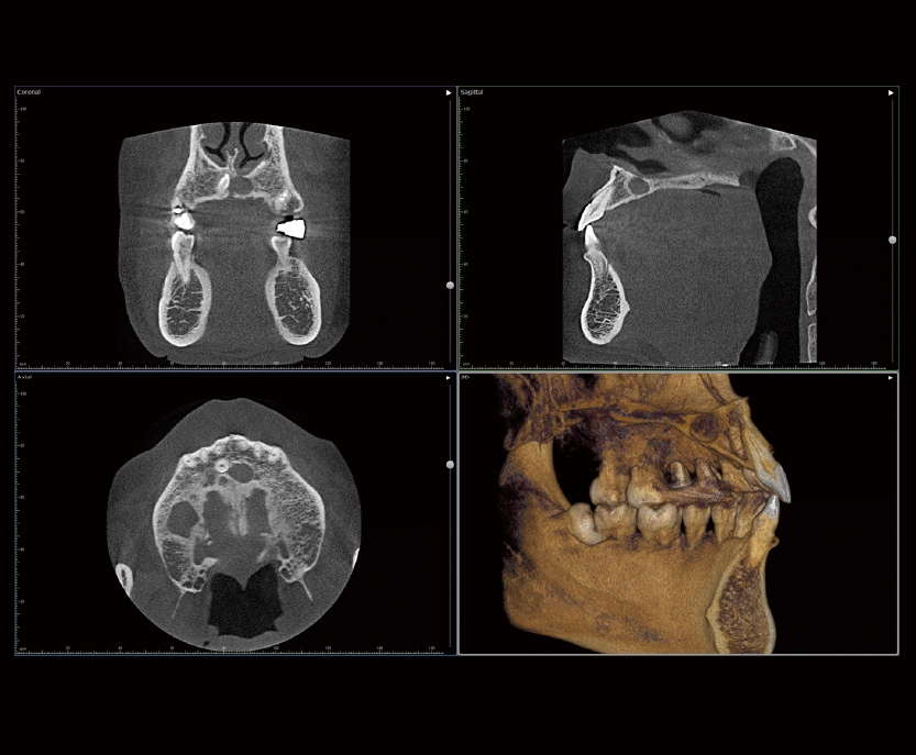
Cyst
One-shot CEPHALO
*SOLIO XZII MAXIM / Specifications with Cephalometric Exposure

76 μm Pixel Size, Giving our Highest Image Quality yet

Equipped with a new cephalometric FPD sensor with a pixel size of
just 76 μm, SOLIO XZII now offers significantly higher resolution.
This makes it possible to obtain more detailed images for easier
diagnosis. Furthermore, the line pair resolution* has also been
improved by approx. 1.7 times compared to the FPD sensors in our
other models.
*Resolution is indicated by the number of black and white line pairs
per mm that can be distinguished.
One-shot Exposure for a Second or Less
With a one-shot exposure time of only 1.0 second maximum, you can
image confidently without being affected by your patient’s body
movement. The shorter exposure time reduces the X-ray dose of
patients, children in particular. Also, there should be no image
blurring as a result of the sensor inadvertently contacting the
patient’s shoulder -which is often an issue during line scanning.
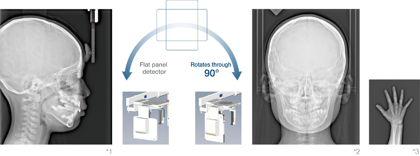
*1 Cephalometric exposure for 1.0 sec (side): with the sensor used
laterally
*2 Cephalometric exposure for 1.0 sec (front): with the sensor oriented vertically
*3 Bone of Carpus for 1.0 sec (Optional)
*2 Cephalometric exposure for 1.0 sec (front): with the sensor oriented vertically
*3 Bone of Carpus for 1.0 sec (Optional)
Comparison with Scanning Method
SOLIO XZII MAXIM
Pixel size: 76 μm (1 sec)
Panoramic X-ray unit our conventional CCD sensor
Pixel size: 96 μm (4 sec)
Comfortable Working Environment
With a variety of functions such as our original Head Support & Grip and
CT Positioning System, a comfortable working environment is assured,
easing the burden on both patient and operator.
User-friendly & Comfortable Setting of the Exposure Area for CT Images
Simple CT operations are possible without burdening the patient or
operator.
Automatic Setting of Image Exposure Area
By selecting your desired image area via the connected PC, the
instrument can automatically move to the image acquisition location.
Prior positioning in the horizontal plane can be performed before
introducing the patient.
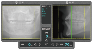
Preliminary Imaging Function
The preliminary imaging function enables directional adjustment of
the CT capture area forwards, backwards, left, right, and up & down.
This greatly assists obtaining an accurate and reliable position
setting. After setting, the CT mechanism automatically moves to the
targeted position for trouble-free image acquisition. This prevents
errors due to incorrect positioning and avoids repetition of CT
imaging operations.
*This video is in Japanese only.
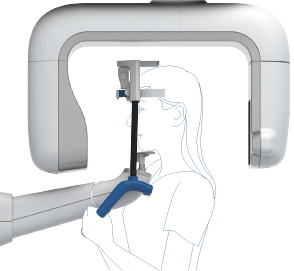
Original Head-support & Grip Assures Correct Positioning for Panoramic Exposures
Total 7-point support including 2 points on the patient’s chest, in
addition to stabilized head positioning. This makes for a steadier
hold on the patient for clearer images with reduced blur. Also
possible is panoramic imaging using the correct extended posture for
the cervical spine. The process of patient position adjustment is
also smooth, thanks to the minimized part-exchanges required for
different exposure modes.
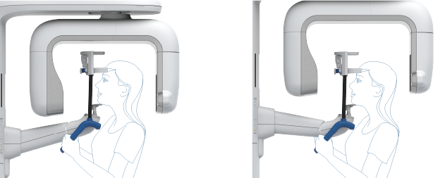
CT Positioning System
After positioning a patient, you can perform vertical movement of
the CT exposure mechanism with a single touch Since it is possible
to adjust the 100 mm wide CT exposure region up or down without
moving the patient, the burden on the patient and the operator is
drastically reduced.
*This video is in Japanese only.
Space-saving, Safe and Secure
Our solutions can be installed to make optimum use of space, for safe
and secure operation over extended periods.
780 mm Stroke for Standing and Sitting Positions
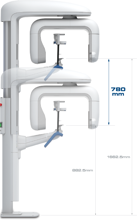
With a vertical stroke width of 780 mm, SOLIO XZII caters for a
wide range of patient positions such as standing, sitting, or
wheelchair bound.
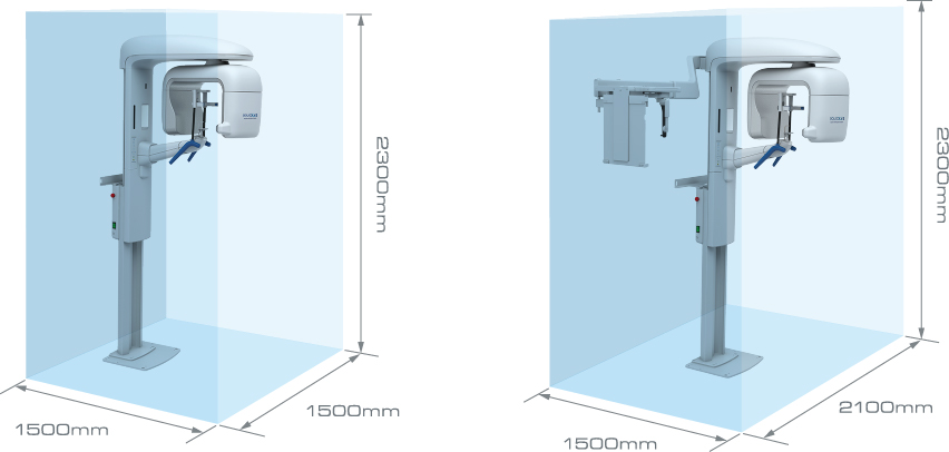
Space-saving and Compact CT Imaging System
SOLIO XZII takes up an area of 1.5 m square, SOLIO XZII MAXIM can be
installed in an X-ray room of 2.1 m width by 1.5 m depth. It is our
most compact CT imaging system yet.
*This video is in Japanese only.
Navigating LED illumination on the Operation Panel
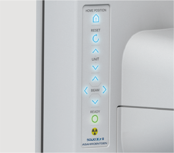
All the necessary operation buttons are incorporated into the
operation panel with a user-friendly, easy-to-see design. With
these buttons you can move the instrument up/down, position the
rotating head to the exposure region, and move the positioning
beam. Moreover, the LED assistance helps you navigate to the
required buttons according to the exposure steps.
Compact Sensor Design and Wide Arm
The space-saving design of the lower section of the sensor and the
wide-arm design minimizes the possibility of the rotating mechanism
contacting the patient’s shoulder during exposure.